Insurance Gas/Electricity Loans Mortgage Attorney Lawyer Donate Conference Call Degree Credit Treatment Software Classes Recovery Trading Rehab Hosting Transfer Cord Blood Claim compensation mesothelioma mesothelioma attorney Houston car accident lawyer moreno valley can you sue a doctor for wrong diagnosis doctorate in security top online doctoral programs in business educational leadership doctoral programs online car accident doctor atlanta car accident doctor atlanta accident attorney rancho Cucamonga truck accident attorney san Antonio ONLINE BUSINESS DEGREE PROGRAMS ACCREDITED online accredited psychology degree masters degree in human resources online public administration masters degree online bitcoin merchant account bitcoin merchant services compare car insurance auto insurance troy mi seo explanation digital marketing degree floridaseo company fitness showrooms stamfordct how to work more efficiently seowordpress tips meaning of seo what is an seo what does an seo do what seo stands for best seotips google seo advice seo steps, The secure cloud-based platform for smart service delivery. Safelink is used by legal, professional and financial services to protect sensitive information, accelerate business processes and increase productivity. Use Safelink to collaborate securely with clients, colleagues and external parties. Safelink has a menu of workspace types with advanced features for dispute resolution, running deals and customised client portal creation. All data is encrypted (at rest and in transit and you retain your own encryption keys. Our titan security framework ensures your data is secure and you even have the option to choose your own data location from Channel Islands, London (UK), Dublin (EU), Australia.
Tendon Diagram / Knee Joint Injections | Arizona Pain Specialists - Phoenix ... : Start studying muscles and tendons.. This important tendon in the back of the calf and ankle stores the elastic energy needed for running, jumping, and other physical activity. Without tendons, your muscles wouldn't be able to make your bones move. The tendons have 2 functions: In the back and elsewhere in the body, tendons attach muscles to bones. Ligaments and tendons serve similar purposes, but in different ways.
This important tendon in the back of the calf and ankle stores the elastic energy needed for running, jumping, and other physical activity. Learn about the anatomy and physiology of tendons. The muscle belly then crosses the entire upper arm and separates into two tendons. Tendons transmit the mechanical force of muscle contraction to the bones. These tendons can become inflamed with overuse activities and from trauma such as ankle sprains.
HanhChampion Blogspot: Basic Arm Exercises from 2.bp.blogspot.com Tendon, tissue that attaches a muscle to other body parts, usually bones. The achilles tendon is the largest. These tendons can become inflamed with overuse activities and from trauma such as ankle sprains. 2 ligaments (trapezoid& conoid ligaments) attach the clavicle coracoid process of scapula these tiny ligaments (w/ acominoclavicular joint) keep scapula attached to clavicle. Allows the action of raising the foot. Start studying muscles and tendons. In the back and elsewhere in the body, tendons attach muscles to bones. Cook and purdum have proposed a new strategy when approaching tendon pain, and this is called the tendon continuum.
Reflex exam (deep tendon reflexes).
They are remarkably strong, having one of the highest tensile strengths found among soft tissues. Related posts of foot tendons and ligaments diagram ankle bones anatomy structure. In the back and elsewhere in the body, tendons attach muscles to bones. They suggest that the tendon can move up and down this. Tendons are thick bands of tissue that connect muscles to bones. Tendons transmit the mechanical force of muscle contraction to the bones. Leg muscle and tendon diagram google search leg muscles anatomy human body anatomy muscle anatomy.the ultrastructure of leg tendon glands shows that the secretory cells are located in three independent regions, separated from each other by unmodified epidermal cells: Tendon, tissue that attaches a muscle to other body parts, usually bones. One tendons inserts onto the forearm bone, the radius, and the second spreads out to join the fascia along the upper part of the forearm. Also allows the action of raising up onto toes. Ligaments and tendons serve similar purposes, but in different ways. Allows the foot to be turned inward and also supports the arch of the foot. Cook and purdum have proposed a new strategy when approaching tendon pain, and this is called the tendon continuum.
We have a collection of human body muscle diagram to help you learn more about the topic. Also allows the action of raising up onto toes. The achilles tendon is the largest. If you would like to learn all the parts of the foot structure, you have come to the right place. Without tendons, your muscles wouldn't be able to make your bones move.
Easy Notes On 【Muscles of the Upper Arm】Learn in Just 3 ... from www.earthslab.com Tendons are thick bands of tissue that connect muscles to bones. 2 ligaments (trapezoid& conoid ligaments) attach the clavicle coracoid process of scapula these tiny ligaments (w/ acominoclavicular joint) keep scapula attached to clavicle. The reactive tendinopathy, tendon disrepair and the degenerative tendinopathy. Start studying muscles and tendons. Brings trunk forward, and aids expiration. A tendon, also known as a sinew, is a fibrous tissue that helps to facilitate this movement. In the back and elsewhere in the body, tendons attach muscles to bones. Related posts of shoulder muscles and tendons diagram muscles of the shoulder and upper.
One of the most important tendons in terms of mobility of the leg is the achilles tendon.
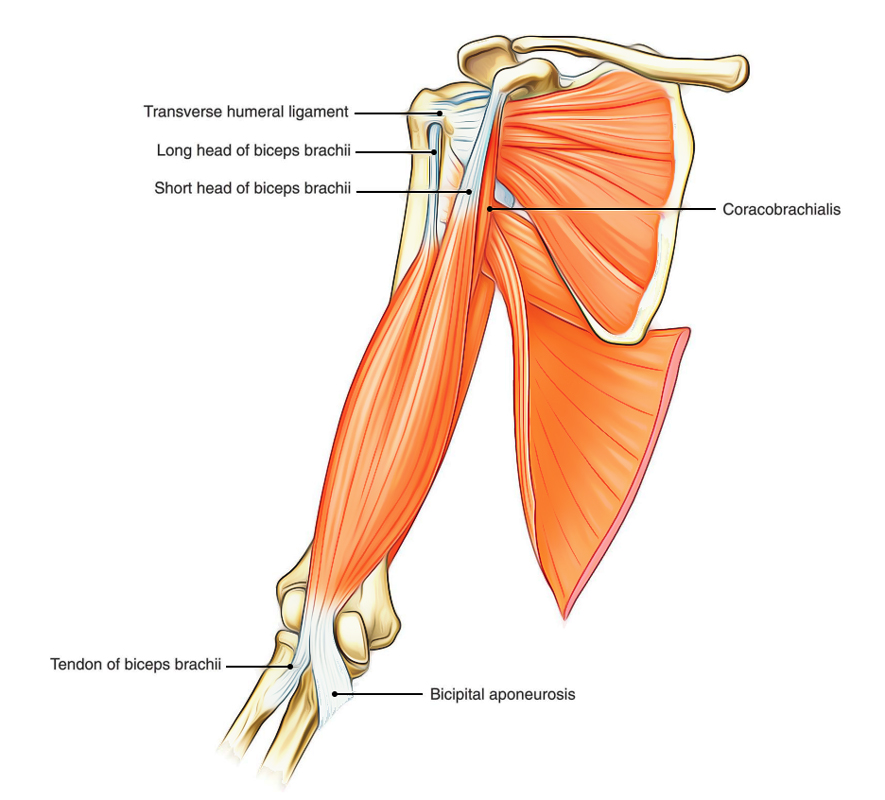
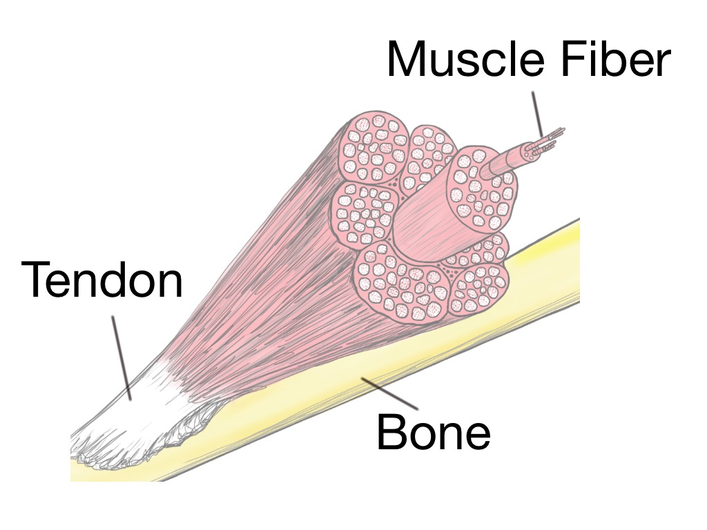
Tendons are the connective tissues between the bones and the muscles. Tendons that make this possible include: Tendons are thick bands of tissue that connect muscles to bones. Posted on april 3, 2019april 3, 2019. The reactive tendinopathy, tendon disrepair and the degenerative tendinopathy. To bend the elbow and to turn the palm of the hand towards the sky. In the back and elsewhere in the body, tendons attach muscles to bones. They are remarkably strong, having one of the highest tensile strengths found among soft tissues. Ligaments join the knee bones and provide stability to the knee: The current term that is recommended to describe this cohort of patients is 'tendinopathy'. Tendons are found throughout the body, from the head and neck all the way down to the feet. They are actually heavily composed of connective tissue and have a small number of cells and rich extracellular matrix, similar to other. Movement occurs when our muscles pull on our bones, relocating them.
Leg muscle and tendon diagram google search leg muscles anatomy human body anatomy muscle anatomy.the ultrastructure of leg tendon glands shows that the secretory cells are located in three independent regions, separated from each other by unmodified epidermal cells: 1 tendons join muscles to their corresponding bones. Cook and purdum have proposed a new strategy when approaching tendon pain, and this is called the tendon continuum. For more anatomy content please follow us and visit our website: Reflex exam (deep tendon reflexes).
Soft Tissue Therapy - Explained - PreHab Exercises from www.prehabexercises.com Tendons that make this possible include: The reactive tendinopathy, tendon disrepair and the degenerative tendinopathy. The fleshy, thick part of the muscle is called its belly. We are pleased to provide you with the picture named right arm muscle and tendon anatomy.we hope this picture right arm muscle and tendon anatomy can help you study and research. Tendons are the connective tissues between the bones and the muscles. Leg muscle and tendon diagram google search leg muscles anatomy human body anatomy muscle anatomy.the ultrastructure of leg tendon glands shows that the secretory cells are located in three independent regions, separated from each other by unmodified epidermal cells: Attaches the calf muscles to the calcaneus, most important muscles for running, jumping, walking etc. It can be used by a teacher or student for academic purposes.
The patellar tendon holds the patella with other two bones, similarly iliotibial band helps in supporting tibia and fibula.
Related posts of foot tendons and ligaments diagram ankle bones anatomy structure. Check out and click on the image to download it. A body muscle diagram is used by different people for various uses. If you would like to learn all the parts of the foot structure, you have come to the right place. The patellar tendon holds the patella with other two bones, similarly iliotibial band helps in supporting tibia and fibula. They propose there are 3 stages to this continuum. Tendons, located at each end of a muscle, attach muscle to bone. Fall on one point of shoulder and can rupture these ligaments with dislocation of ac joint. By cliparea l custom media. Tendons generally have a very complex structure; The tendons have 2 functions: In the back and elsewhere in the body, tendons attach muscles to bones. The achilles tendon is the largest.